Imaging Case 21:
Prostate cancer - Adenocarcinoma
To effectively plan and guide a prostatectomy, surgeons rely on preoperative imaging, such as MRI and PET-CT. This imaging plays a crucial role in selecting the optimal surgical approach and assessing the potential for nerve preservation. MRI, in particular, provides detailed visualization of nerves, blood vessels, and the prostatic capsule, helping surgeons decide whether to preserve or sacrifice the neurovascular bundle—a critical factor influencing postoperative quality of life.
During radical prostatectomy, removing the neurovascular bundle may be necessary if cancer has infiltrated these structures, which can result in erectile dysfunction and urinary incontinence. A nerve-sparing approach aims to preserve sexual function and continence but increases the risk of incomplete tumor removal if cancer extends beyond the visible tumor margins. Since preoperative imaging does not always accurately define the full extent of a tumor, incorporating intraoperative information can help ensure a more complete resection. Recent studies have demonstrated that high-resolution specimen PET-CT imaging using PSMA-based radiotracers provides a precise visualization of the resected prostate tumor, potentially aiding in the assessment of resection completeness [1,3,4,5].
In this case, we demonstrate how intraoperative specimen PET-CT imaging with 18F-PSMA can effectively visualize tumors within resected tissue specimens. By harnessing high resolution and enhanced accuracy, these images have the potential to offer greater precision than the current preoperative imaging. This real-time intraoperative information can optimally guide surgeons in decision-making during the procedure, potentially leading to improved clinical outcomes.
This case is presented with the support of dr. B. Lambert, Prof. dr. K. Decaestecker, dr. F. Ameye, dr. P. De Backer, and colleagues of AZ Maria Middelares hospital, Ghent, Belgium, and is part of the investigator driven study to explore potential indications of an intraoperative specimen PET-CT imager (Trial registration number: BUN: B0172022000009).

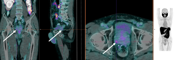 Figure 1. Preoperative 18F -PSMA PET-CT image. High uptake is shown in a small focus posterolaterally on the right, with SUVmax of 3.5.
Patient history
A 73-year-old patient was diagnosed with invasive prostatic adenocarcinoma (Gleason score 7). Preoperative 18F-PSMA PET-CT imaging showed a small focus in the right posterolateral zone, see Fig. 1. However, preoperative MRI imaging showed a significant lesion in the peripheral zone of the left apex. Additionally, small lesions were observed bilaterally in the mid-gland peripheral zone on MRI.
Biopsy confirmed invasive adenocarcinoma, with Gleason score 4+3 in the right region and 3+4 in the left region of the prostate. The patient was scheduled for robot assisted radical prostatectomy.
Specimen PET-CT imaging
The patient was intravenously injected with 1 MBq/kg of 18F-PSMA during surgery.
Resection of the prostate specimen was completed at approximately 173 min after injection. Immediately after resection, a high-resolution specimen PET-CT image was acquired in the operating theatre. The images are shown in Fig. 2. In this image three orthogonal views of the specimen PET-CT images are shown. The PET images are represented in color scale, superimposed on the CT images in greyscale. The images of the prostate specimen clearly show increased 18F-PSMA uptake with SUVmax of 16. Moreover, at the right (1) and left (2) side of the specimen, 18F-PSMA uptake is seen at a certain distance of the border of the specimen.
Figure 2. Transverse, coronal, and sagittal slices and 3D view of the specimen PET-CT images of the prostate specimen. Specimen orientation is as indicated. The tumor is represented by a bright colorful region in the specimen. The PET tracer scale bar is depicted on the right-hand side. At the right (1) and left (2) side of the specimen, 18F-PSMA uptake is seen at a certain distance of the border of the specimen.
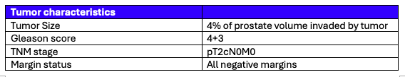 Table 1. Tumor characteristics assessed by histopathological evaluation.
Histopathological evaluation
After specimen PET-CT imaging, the surgical specimen was sent to the pathology department for routine histopathological evaluation, which was available after several days. The histopathological results are listed in Table 1.
Two lesions were seen at the left and right side of the prostate, for which all surgical margins were found negative. This is visualized in Figure 3. This correlates excellently with the regions of radiotracer uptake on the specimen PET-CT image.
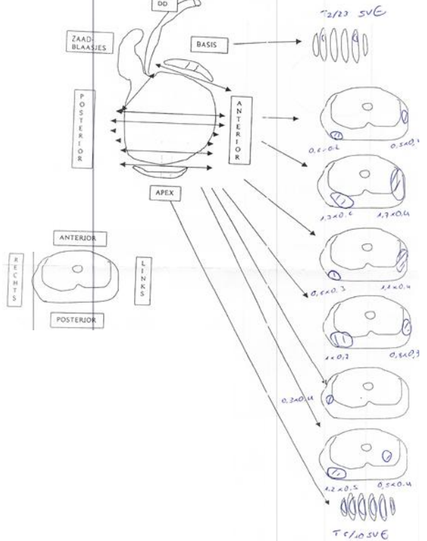 Figure 3. A drawing of the histopathological results.
Discussion and conclusion
Preoperative MRI and PET-CT guide prostate cancer surgery, but when tumor sites are missed, the risk of incomplete removal and recurrence increases.
In this case, specimen PET-CT provided unmatched precision, aligning with histopathology and preoperative imaging. While PET-CT detected uptake on the right and MRI identified a left-sided lesion, specimen PET-CT successfully visualized both, with histopathology confirming their presence. Additionally, a highlighted radiotracer uptake near – but not touching - the border of the specimen was verified by histopathology as a negative surgical margin, confirming clean resection margins.
This case demonstrates the value of intraoperative specimen PET-CT —delivering real-time, tumor visualization beyond preoperative imaging. With immediate surgical feedback, this technology may enhance precision, help minimize recurrence risk, and can therefore help improve patient outcomes—empowering surgeons with the confidence to achieve the best possible results.
References
[1] Darr C et al. (2023). Intraoperative Molecular Positron Emission Tomography Imaging for Intraoperative Assessment of Radical Prostatectomy Specimens. European Urology Open Science 54:28-32
[2] Okarvi, S. M. et al. (2019). Recent developments of prostate-specific membrane antigen (PSMA)-specific radiopharmaceuticals for precise imaging and therapy of prostate cancer: an overview. Clin. Transl. Imaging 7:189–208. issn: 22817565
[3] Muraglia L et al. (2023). First Live-Experience Session with PET/CT Specimen Imager: A Pilot Analysis in Prostate Cancer and Neuroendocrine Tumor. Biomedicines 11:645.
[4] Lambert B. et al. (2025) Feasibility study on the implementation of a mobile high-resolution PET/CT scanner for surgical specimens: exploring clinical applications and practical considerations. Eur J Nucl Med Mol Imaging. https://doi.org/10.1007/s00259-025-07143-z
[5] Moraitis A. et al. (2025) Evaluation of Surgical Margins with Intraoperative PSMA PET/CT and Their Prognostic Value in Radical Prostatectomy. jnumed. 124.268719. https://doi.org/10.2967/jnumed.124.268719