Imaging Case 20:
Prostate cancer - Adenocarcinoma
To effectively plan and guide a prostatectomy, surgeons rely on preoperative imaging, such as MRI and PET-CT, to precisely determine the location, size, and extent of tumors, helping them choose the optimal surgical approach. Additionally MRI provides detailed visualization of nerves and blood vessels, aiding in the decision of whether to preserve or sacrifice the neurovascular bundle—a key factor in post-surgical quality of life.
In cases of radical prostatectomy, the neurovascular bundle may need to be sacrificed, potentially leading to erectile dysfunction and loss of continence. A nerve-sparing approach can reduce these risks but comes with an increased chance of incomplete tumor removal. Therefore, accurate preoperative imaging is essential to balance the goals of preserving healthy tissue while ensuring complete tumor resection.
Important to note is that pelvic lymph node dissection is often performed during prostate cancer surgery, with the aim of obtaining detailed prognostic information as well as removing metastatically invaded lymph nodes detected on preoperative imaging. However, removal of all suspicious lymph nodes during surgery can be challenging, for which the surgeon has no information at the point of surgery. Recent publications have shown that high-resolution specimen PET-CT imaging may provide such insights [1,3,4,5].
In this case, we demonstrate how intraoperative specimen PET-CT imaging with 18F-PSMA can effectively visualize tumors within resected tissue specimens. By harnessing high resolution and enhanced accuracy, these images have the potential to offer greater precision than the current preoperative imaging. This real-time intraoperative information can optimally guide surgeons in decision-making during the procedure, potentially leading to improved clinical outcomes.
This case is presented with the support of dr. B. Lambert, Prof. dr. K. Decaestecker, dr. F. Ameye, dr. P. De Backer, and colleagues of AZ Maria Middelares hospital, Ghent, Belgium, and is part of the investigator driven study to explore potential indications of an intraoperative specimen PET-CT imager (Trial registration number: BUN: B0172022000009).
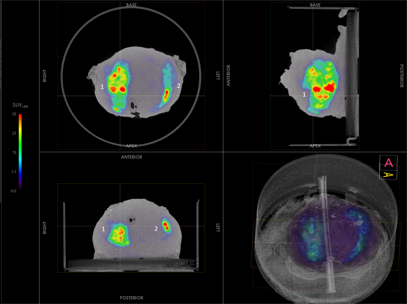
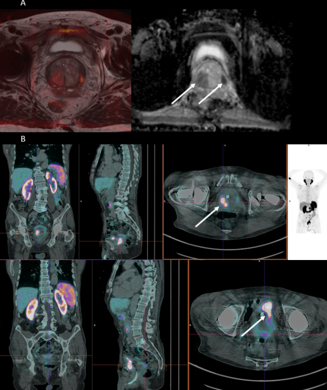 Figure 1. (A) Preoperative MRI images, showing bilateral invasion. (B) Preoperative 18F -PSMA PET-CT images with high uptake at the left side (SUVmax 11.19) and right side (SUVmax 29.40).
Patient history
Specimen PET-CT Imaging - prostate specimen
The patient was intravenously injected with 1 MBq/kg of 18F-PSMA at the start of the surgery.
Resection of the prostate specimen was completed at 111 min after injection. Immediately after resection, a high-resolution specimen PET-CT image was acquired in the operating theatre. The images are shown in Fig. 2. In this image three orthogonal views of the specimen PET-CT images are shown. The PET images are represented in color scale, superimposed on the CT images in greyscale. The images of the prostate specimen clearly show increased 18F-PSMA uptake on both sides of the prostate with SUVmax of 30.6. Bilaterally, the 18F-PSMA uptake is seen at a clear distance from the border of the specimen and suggests there is no extracapsular invasion.
Figure 2: Transverse, coronal, and sagittal slices and 3D view of the specimen PET-CT images of the prostate specimen. Specimen orientation is as indicated. The tumor is represented by a bright colorful region in the specimen. The PET tracer scale bar is depicted on the right-hand side. Both on the right (1) and on the left (2) side of the specimen, 18F-PSMA uptake is seen. This uptake is at a certain distance from the border of the specimen.
Specimen PET-CT Imaging - lymph nodes
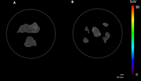 Figure 3: Specimen PET-CT images of resected pelvic lymph nodes, distributed over two specimen containers (A-B). Moreover, (A) two lymph nodes of the right side, and (B) Five lymph nodes of the left side. The PET tracer scale bar is depicted on the right-hand side. No significant 18F-PSMA uptake is visualized in all lymph nodes.
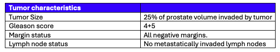 Table 1. Tumor and lymph node characteristics assessed by histopathological evaluation.
Histopathological evaluation
After specimen PET-CT imaging, the surgical specimen was sent to the pathology department for routine histopathological evaluation, which was available after several days. The histopathological results are listed in Table 1.
All surgical margins were found negative, and contrary to what was suspected from preoperative imaging there was no extracapsular invasion. All lymph nodes showed no evidence of metastases. This correlates excellently with the regions of radiotracer uptake on the specimen PET-CT images.
Discussion and conclusion
Radical prostatectomy demands a careful balance—removing cancer while preserving nerve structures vital to quality of life. A nerve-sparing approach reduces the risk of impotence and incontinence but makes achieving clear surgical margins more challenging. To navigate this, surgeons rely on preoperative MRI and PET-CT to map tumor size, location, and extent.
In this case, specimen PET-CT identified two uptake regions—one on the left and one on the right—for which the uptake was seen at a distance of the specimen border. Histopathology confirmed the presence of the two lesions, both assessed as negative, further validating the accuracy of this technique. Notably, while preoperative imaging suggested capsular invasion on the right, this finding was not supported by specimen PET-CT, nor histopathology.
With intraoperative specimen PET-CT, surgeons gain real-time insights, enabling more informed decision-making during surgery. This innovative imaging approach may help optimize cancer removal, reduce positive margins, and improve patient outcomes.
References
[1] Darr C et al. (2023). Intraoperative Molecular Positron Emission Tomography Imaging for Intraoperative Assessment of Radical Prostatectomy Specimens. European Urology Open Science 54:28-32
[2] Okarvi, S. M. et al. (2019). Recent developments of prostate-specific membrane antigen (PSMA)-specific radiopharmaceuticals for precise imaging and therapy of prostate cancer: an overview. Clin. Transl. Imaging 7:189–208. issn: 22817565
[3] Muraglia L et al. (2023). First Live-Experience Session with PET/CT Specimen Imager: A Pilot Analysis in Prostate Cancer and Neuroendocrine Tumor. Biomedicines 11:645.
[4] Lambert B. et al. (2025) Feasibility study on the implementation of a mobile high-resolution PET/CT scanner for surgical specimens: exploring clinical applications and practical considerations. Eur J Nucl Med Mol Imaging. https://doi.org/10.1007/s00259-025-07143-z
[5] Moraitis A. et al. (2025) Evaluation of Surgical Margins with Intraoperative PSMA PET/CT and Their Prognostic Value in Radical Prostatectomy. jnumed. 124.268719. https://doi.org/10.2967/jnumed.124.268719