Imaging Case 18:
Prostate cancer - Adenocarcinoma
To guide and plan a prostatectomy, the surgeon relies on preoperative imaging. For example, MRI and PET-CT help identify the exact location, size, and extent of tumors, allowing the surgeon to decide on the best approach. MRI can visualize nerves and blood vessels, which is used to determine whether the neurovascular bundle needs to be sacrificed or not. Radical prostatectomy may require sacrificing the neurovascular bundle, which impacts the quality of life of prostate cancer patients. A nerve-sparing approach can be used to reduce postoperative impotence and incontinence, but this involves an increased risk of incomplete resection. As preoperative imaging can’t always accurately predict the extent of the tumor, the addition of intraoperative information may be useful to ensure completeness of resection. Recent publications have shown that high-resolution specimen PET-CT imaging with PSMA-based radiotracers can provide an accurate view of the resected prostate tumor and may help enable assessing completeness of resection [1,3,4,5].
In this case, we demonstrate how intraoperative specimen PET-CT imaging with 18F-PSMA can effectively visualize tumors within resected tissue specimens. By harnessing high resolution and enhanced accuracy, these images have the potential to offer greater precision than the current preoperative imaging. This real-time intraoperative information can optimally guide surgeons in decision-making during the procedure, potentially leading to improved clinical outcomes.
This case is presented with the support of dr. B. Lambert, Prof. dr. K. Decaestecker, dr. F. Ameye, dr. P. De Backer, and colleagues of AZ Maria Middelares hospital, Ghent, Belgium, and is part of the investigator driven study to explore potential indications of an intraoperative specimen PET-CT imager (Trial registration number: BUN: B0172022000009).
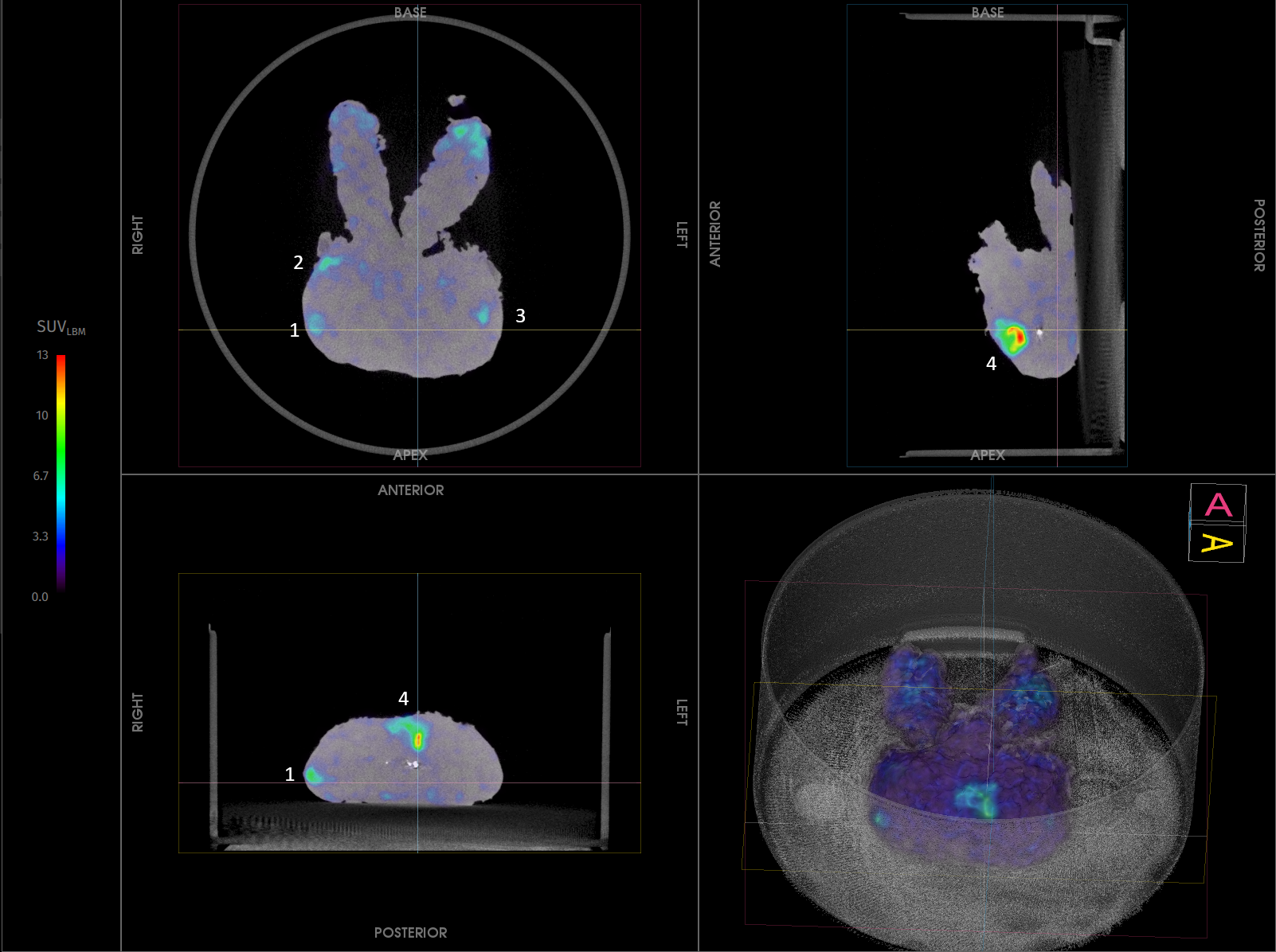
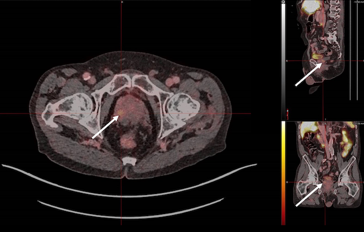 Figure 1. Preoperative 18F -PSMA PET-CT image, acquired one week before surgery. High uptake is shown at the base of the prostate in the right peripheral zone, with SUVmax 5.53.
Patient History
A 75-year-old patient was diagnosed with prostatic acinar adenocarcinoma (Gleason score 4+3). An MRI was performed twenty-nine weeks prior to surgery, showing a lesion on the left side of the prostate. Remarkably, a preoperative 18F -PSMA PET-CT scan, performed one week prior to surgery, identified a solitary lesion at the base of the prostate in the right peripheral zone (SUVmax 5.53), see Fig. 1.
Biopsy confirmed acinar adenocarcinoma, with highest and most frequent Gleason score 4+3 at the left anterior zone and left posterior zone. On the right side, limited involvement with Gleason score 3+3 was noted.
It is important to highlight that while the MRI indicated a suspicious lesion on the left side of the prostate and a biopsy confirmed malignancy in this area, the lesion did not exhibit PSMA positivity on the PSMA PET-CT scan, which showed uptake only on the right side.
The patient was staged T2u (PRIMARY 4), N0M0, according to miTNM classification and scheduled for robot-assisted radical prostatectomy (single port).
Specimen PET-CT images
The patient was intravenously injected with 1 MBq/kg of 18F-PSMA at the start of the surgery.
Resection of the prostate specimen was completed at 140 min after injection. Immediately after resection, a high-resolution specimen PET-CT image was acquired in the operating theatre. The images are shown in Fig. 2. In this image three orthogonal views of the specimen PET-CT images are shown. The PET images are represented in color scale, superimposed on the CT images in greyscale.
The images of the prostate specimen clearly show increased 18F-PSMA uptake with SUVmax of 13, see Fig. 2. Moreover, at the right side of the PET-CT image, two avid foci were detected (1,2) close to but not reaching the border of the specimen, as seen on the preoperative PET-CT images, and at the left side of the prostate, a focus was detected at a small distance from the border of the specimen (3), in correlation to the MRI. At the apical anterior zone of the prostate, a clear uptake pattern was observed as well touching the border of the specimen (4), which was not seen on preoperative imaging.
Figure 2. Transverse, coronal, and sagittal slices and 3D view of the specimen PET-CT images of the prostate specimen. Specimen orientation is as indicated. The tumor is represented by a bright colorful region in the specimen. The PET tracer scale bar is depicted on the right-hand side. At the right side of the PET-CT image, two 18F-PSMA avid foci were detected (1,2), as seen on the preoperative PET-CT images, and at the left side of the prostate, a 18F-PSMA avid focus was detected (3), in correlation to the MRI. At the apical anterior zone of the prostate, a clear uptake pattern was observed as well (4).
 Table 1. Tumor characteristics assessed by histopathological evaluation.
Histopathological evaluation
After specimen PET-CT imaging, the surgical specimen was sent to the pathology department for routine histopathological evaluation, which was available after several days. The histopathological results are listed in Table 1.
A positive surgical margin was found at the at the apical anterior central zone (4 mm) and close margin at the right side (0.1 mm) of the specimen. Also at the right side, two tumor foci were detected, and one lesion at the left side, all with negative margins. This is visualized in Fig. 3. These tumor lesions correlate with the regions of high radiotracer uptake on the specimen PET-CT image.
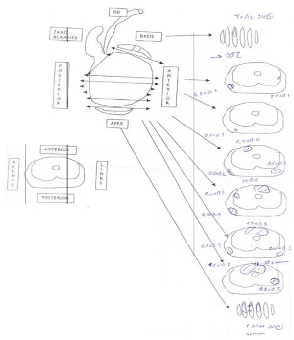 Figure 3. A drawing of the histopathological results.
Discussion and conclusion
A patient diagnosed with prostatic acinar adenocarcinoma presented a unique challenge: preoperative MRI revealed a suspicious lesion on the left side of the prostate, while 18F-PSMA PET-CT showed uptake only on the right side.
The intraoperative PET-CT images of the prostate specimen clearly show increased 18F-PSMA uptake in four regions: At the right side of the PET-CT image two avid foci were detected (1,2), as seen on the preoperative PET-CT imaging, and at the left side of the prostate (3), a focus was detected, in correlation to the MRI. At the apex anterior zone of the prostate, a clear uptake pattern was observed as well (4), which had no correlation with preoperative imaging.
Histopathology validated all four regions as true tumor lesions, proving that intraoperative specimen PET-CT provides a level of tumor visualization beyond preoperative imaging alone. In fact, the lesion that was only visualized on specimen PET-CT imaging turned out to be the lesion with a positive surgical margin.
Surgeons rely on preoperative MRI and PET-CT to map out the size, location, and extent of prostate tumors before surgery. However, when imaging misses critical tumor sites, it increases the risk of incomplete tumor removal, potentially leading to recurrence.
With intraoperative specimen PET-CT, surgeons gain real-time insights into tumor distribution in the excised tissue, helping them make informed surgical decisions on the spot. This advanced imaging technique can enhance oncological outcomes, reduce the risk of positive margins, and improve patient prognosis.
References
[1] Darr C et al. (2023). Intraoperative Molecular Positron Emission Tomography Imaging for Intraoperative Assessment of Radical Prostatectomy Specimens. European Urology Open Science 54:28-32
[2] Okarvi, S. M. et al. (2019). Recent developments of prostate-specific membrane antigen (PSMA)-specific radiopharmaceuticals for precise imaging and therapy of prostate cancer: an overview. Clin. Transl. Imaging 7:189–208. issn: 22817565
[3] Muraglia L et al. (2023). First Live-Experience Session with PET/CT Specimen Imager: A Pilot Analysis in Prostate Cancer and Neuroendocrine Tumor. Biomedicines 11:645.
[4] Lambert B. et al. (2025) Feasibility study on the implementation of a mobile high-resolution PET/CT scanner for surgical specimens: exploring clinical applications and practical considerations. Eur J Nucl Med Mol Imaging. https://doi.org/10.1007/s00259-025-07143-z
[5] Moraitis A. et al. (2025) Evaluation of Surgical Margins with Intraoperative PSMA PET/CT and Their Prognostic Value in Radical Prostatectomy. jnumed. 124.268719. https://doi.org/10.2967/jnumed.124.268719