Precisely where it matters
In surgical oncology, such as breast conserving surgery, rapid assessment of the excised specimen is of utmost importance. You want to be confident that your surgery reached its goal of resecting the target lesion, so that your patients recovery can be promoted.
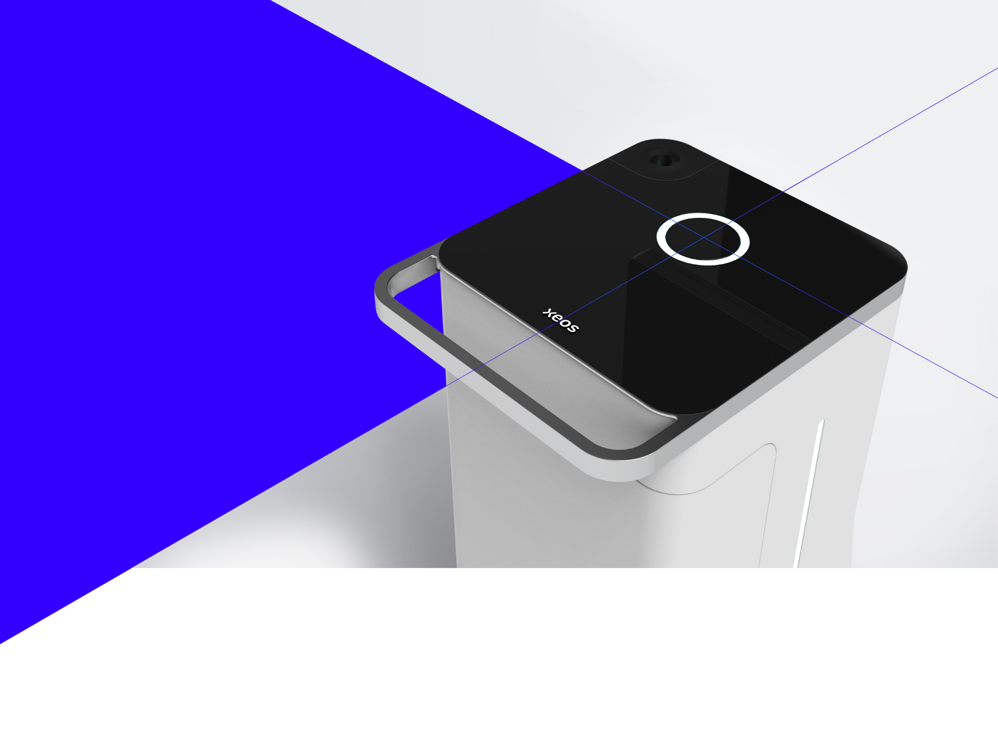
Discover AURA 10High-quality medical grade display monitor
Can be tilted and swiveled to give you more viewing comfort.Unique specimen container handling
Motorized top-load specimen receptacle.Curved transportation handle
Provides intuitive handling, allowing you to fully focus on your patient.Colored LED bars
Show when the x-ray acquisition is taking place and inform you about the progress of the image acquisition.Easy brake system
Allows the unit to be placed securely in the OR.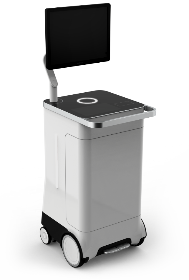

The first PET-CT specimen imager for the OR
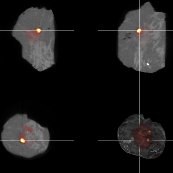
The XEOS AURA 10 is the first-ever PET-CT Specimen Imager for the OR, offering surgeons and imaging specialists the sensitivity of PET imaging, combined with submillimeter spatial resolution.
AURA 10 has made molecular imaging available in the OR. As a result, surgeons and imaging specialists have never been able to perform specimen imaging with so much confidence. Ultimately, patients will be the ones who benefit.
Discover how AURA 10 creates impact for your patientsMolecular imaging
in an instance
Molecular imaging has been the gold standard in oncology for years. Whole-body PET-CT scanners have proven to be invaluable for the diagnosis and follow up of cancers, because they provide unparalleled sensitivity for the detection and localization of malignant cells.
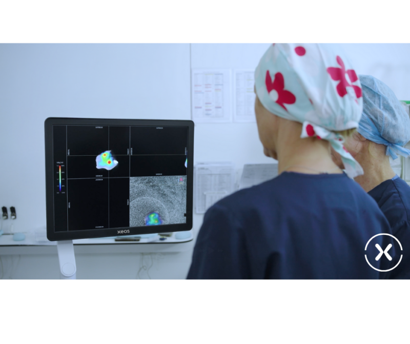
Imagine you could obtain this level of precision within 10 minutes after excision, right at the point of surgery, without having to transport your specimen over to the radiology department. Imagine how this could boost your surgical confidence and improve the wellbeing of your patients.
Discover the benefits
